
")
")

")
")
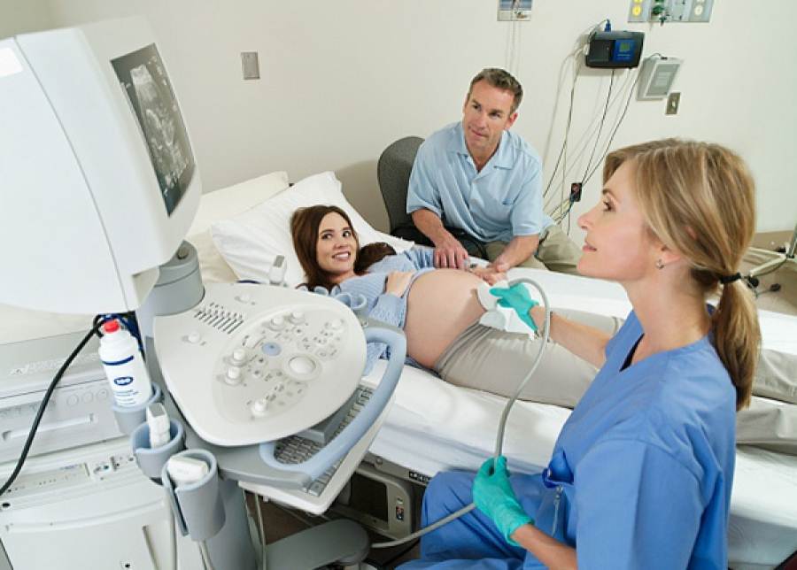
“GENESIS ATHENS” has a well-organised department providing General and Gynaecological Ultrasound Scans. We use state-of-the-art ultrasound tomographs, 3D/4D colour imaging and specialised scientific staff (gynaecologists-radiologists) to perform ultrasound scans of all organs of the human body. Our gynecologists use the cervical slide ultrasound to monitor the health of both the pregnant woman and foetus in the first quarter of pregnancy. In the second quarter of pregnancy, we can perform a comprehensive foetal anatomy ultrasound scan (level II) for a detailed study of foetal anatomy. In the last quarter of pregnancy, a colour Doppler ultrasound can be used to monitor the proper development and growth of the foetus. Further, the Clinic can carry out invasive procedures during pregnancy, such as chorionic villi sampling to screen the chorionic villi (placenta), and amniotic fluid sampling –amniocentesis (between weeks 16 and 20). In addition, this department is responsible for the timely, painless and accurate detection of disorders in other organs of the body, such as the upper and lower abdomen, scrotum, soft tissues, thyroid, breasts, and also performs vascular colour ultrasound scans (triplex).
Mammograph
There is near-universal agreement that prevention is preferable to treatment of disease. If there is no problem why would someone undergo examinations? However, since breast disease appears with increasing frequency, all women should undergo relevant examinations.
It is necessary to have a clinical breast examination for cancer performed by a trained health professional as part of a preventive and diagnostic mammography program, to allow the management of any identified problem at an early stage. The sonner, the better.
When should a woman start having clinical breast examinations? A screening mammogram is performed as a first index test around the age of 35, in case of a positive family history, and should be repeated as a routine examination by all woman after the age of 40, every 1 to 2 years.
Before the age of 35, it is usually sufficient to start around the age of 25 with a breast examination every year and a high-definition ultrasound screening, if necessary.
Don’t let yourself hear the devastating words “…if only you had come sooner…”, have yourself checked NOW.
We, at the “GENESIS ATHENS” Clinic, with our qualified staff and the high-definition and low-radiation, top-of-the-line mammograph, are here to help in the timely prevention of breast cancer.
Medical imaging laboratories
X- ray department
The x-ray laboratory of “GENESIS ATHENS” is equipped with a high-end, low-dosage x-ray machine which can perform a comprehensive range of examinations, from simple radiographs to x-ray radioscopy, pyelograms, cystograms and stomach examination of the large intestine. In addition, it performs painless hysterosapingograms to determine the cause of infertility. X-ray dye is injected through a special balloon catheter to picture the cervical channel, uterine cavity and Fallopian tubes. This technique is used under X-ray radioscopy, lasts 10-15 minutes and recovery is painless. It reveals uterine abnormalities, detects any defects related to polyps or submucous fibroids, determines if the tubes are patent and free of blockage, and identifies the presence of hydrosalpinx and peritubal adhesions.